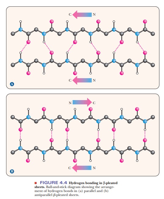
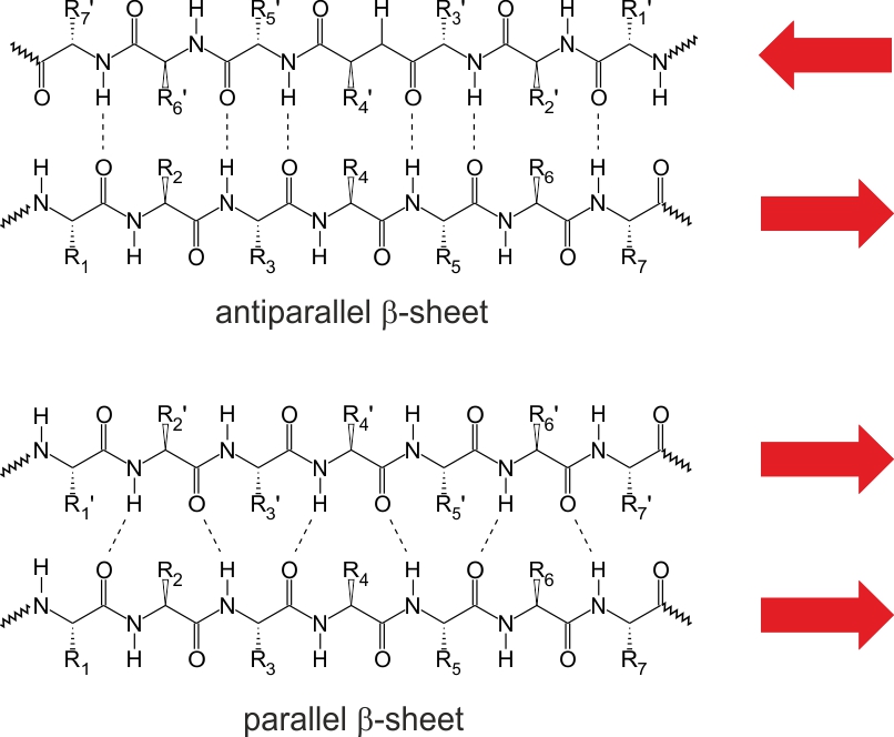
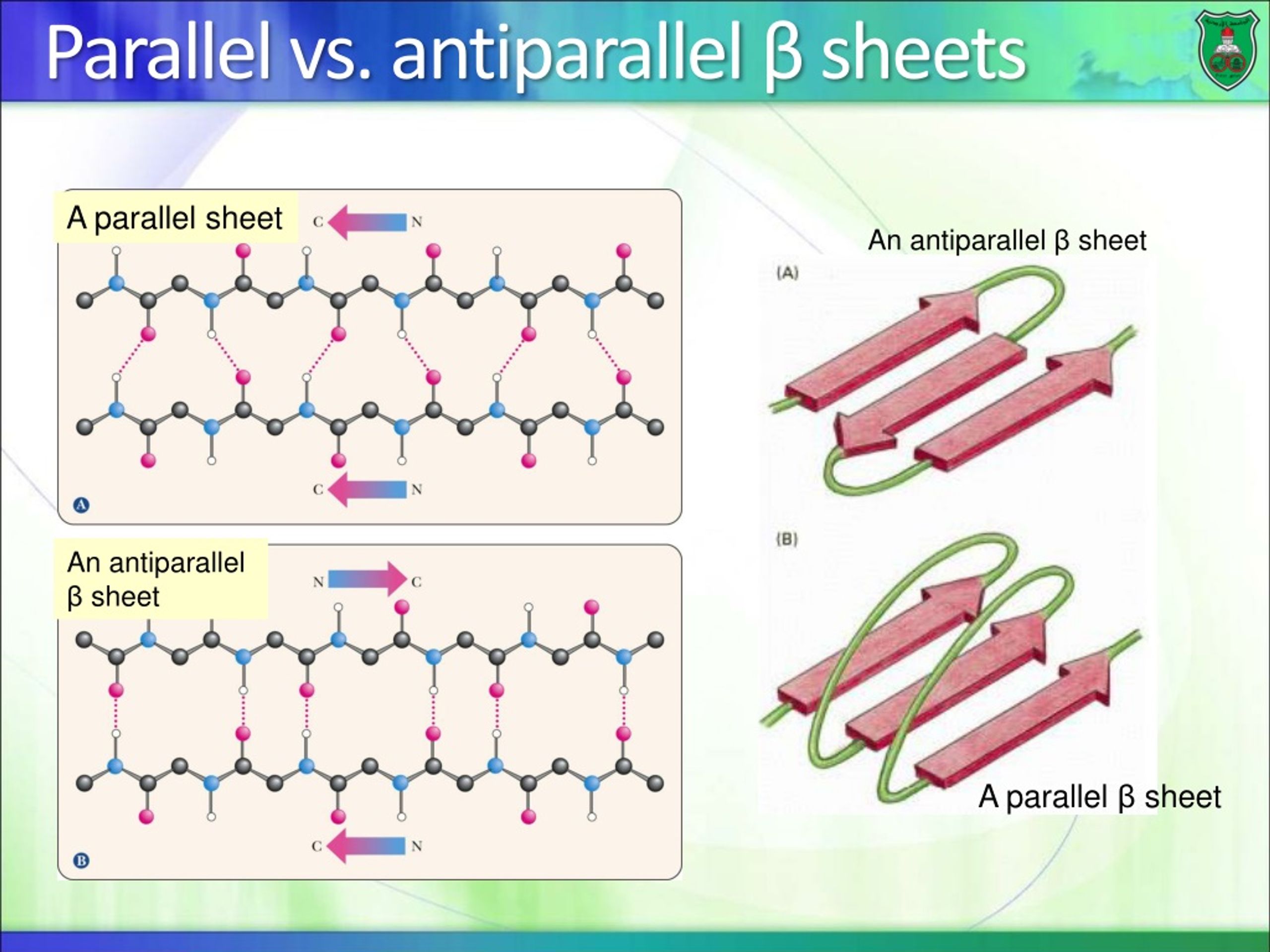
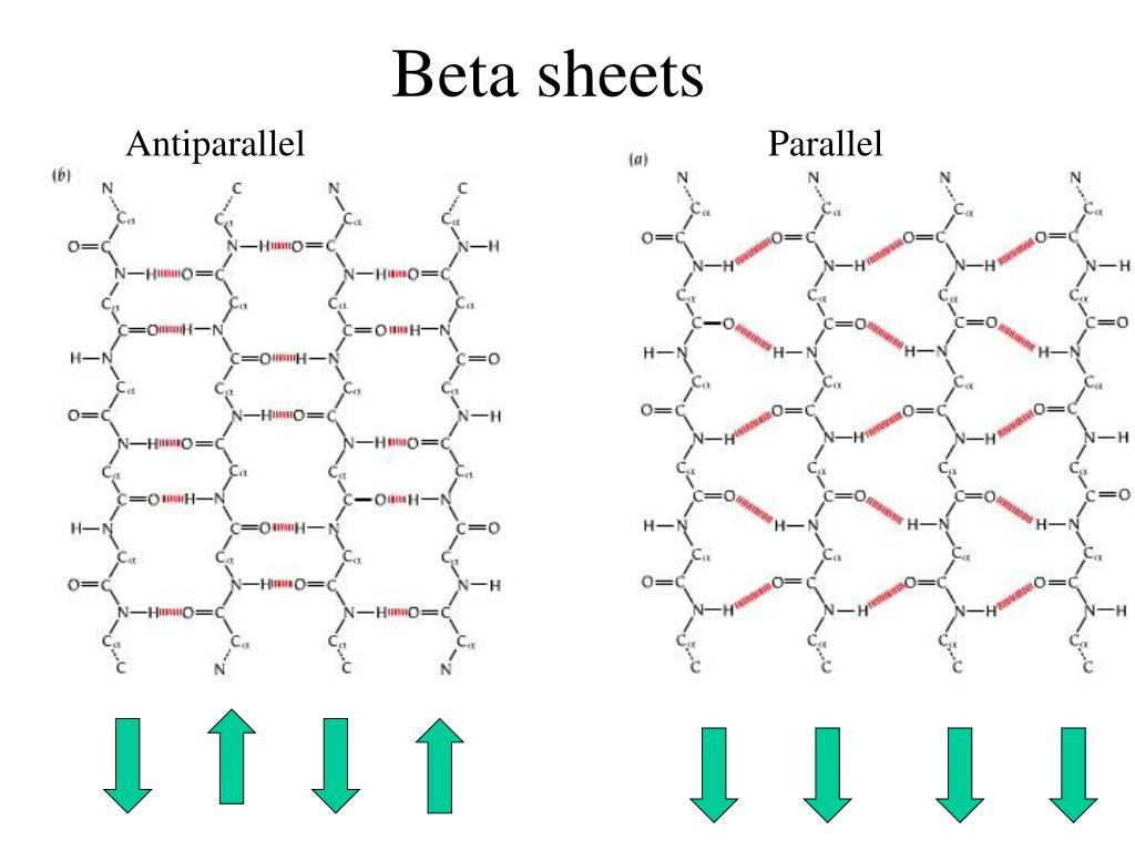
Parallel Beta Sheet Vs Antiparallel - Antiparallel beta pleated sheets and parallel beta pleated sheets are two common structural motifs found in proteins. Antiparallel and parallel β sheets are key secondary structures in proteins. Antiparallel β sheets have strands aligned in opposite. Distinguish between parallel and antiparallel beta strands, and describe how hydrogen bonding patterns and dihedral angles contribute to the.
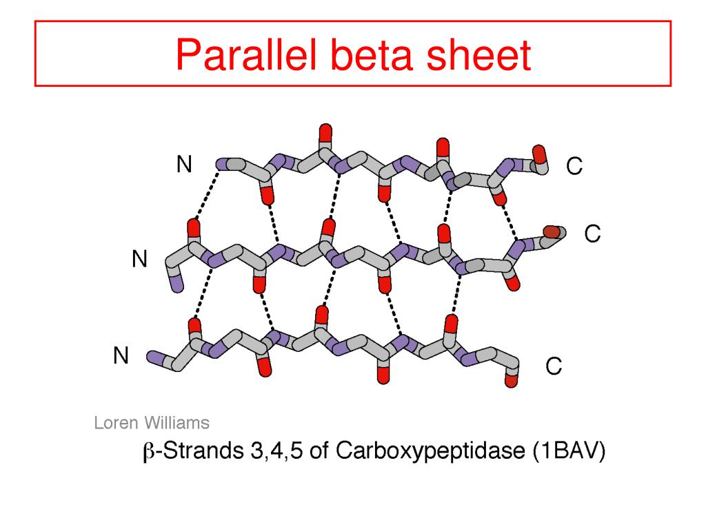
Antiparallel β sheets have strands aligned in opposite. Antiparallel beta pleated sheets and parallel beta pleated sheets are two common structural motifs found in proteins. Distinguish between parallel and antiparallel beta strands, and describe how hydrogen bonding patterns and dihedral angles contribute to the. Antiparallel and parallel β sheets are key secondary structures in proteins.
Antiparallel beta pleated sheets and parallel beta pleated sheets are two common structural motifs found in proteins. Distinguish between parallel and antiparallel beta strands, and describe how hydrogen bonding patterns and dihedral angles contribute to the. Antiparallel and parallel β sheets are key secondary structures in proteins. Antiparallel β sheets have strands aligned in opposite.

Parallel vs antiparallel betasheets YouTube
Antiparallel and parallel β sheets are key secondary structures in proteins. Antiparallel β sheets have strands aligned in opposite. Distinguish between parallel and antiparallel beta strands, and describe how hydrogen bonding patterns and dihedral angles contribute to the. Antiparallel beta pleated sheets and parallel beta pleated sheets are two common structural motifs found in proteins.
PPT Levels of Protein Structure PowerPoint Presentation, free
Antiparallel and parallel β sheets are key secondary structures in proteins. Antiparallel beta pleated sheets and parallel beta pleated sheets are two common structural motifs found in proteins. Distinguish between parallel and antiparallel beta strands, and describe how hydrogen bonding patterns and dihedral angles contribute to the. Antiparallel β sheets have strands aligned in opposite.
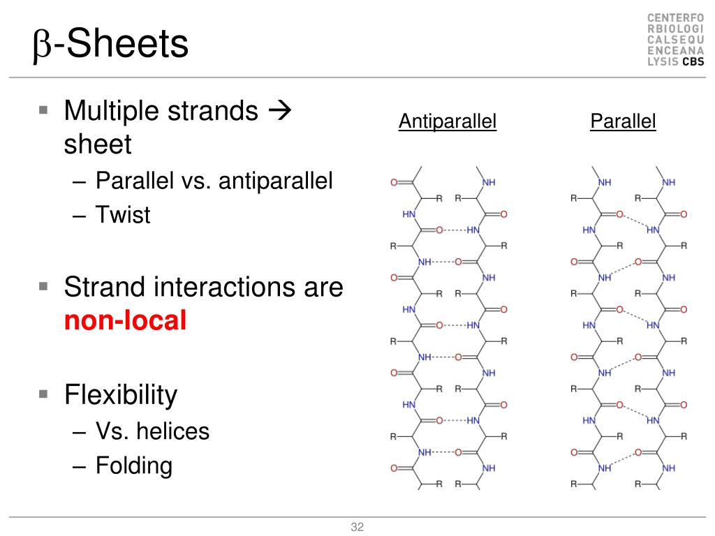
PPT Molecular Biophysics Lecture 2 Protein Structure II PowerPoint
Distinguish between parallel and antiparallel beta strands, and describe how hydrogen bonding patterns and dihedral angles contribute to the. Antiparallel and parallel β sheets are key secondary structures in proteins. Antiparallel β sheets have strands aligned in opposite. Antiparallel beta pleated sheets and parallel beta pleated sheets are two common structural motifs found in proteins.
Beta Pleated Sheet Parallel Vs Antiparallel
Antiparallel β sheets have strands aligned in opposite. Antiparallel and parallel β sheets are key secondary structures in proteins. Antiparallel beta pleated sheets and parallel beta pleated sheets are two common structural motifs found in proteins. Distinguish between parallel and antiparallel beta strands, and describe how hydrogen bonding patterns and dihedral angles contribute to the.
20.15 Secondary Protein Structure Chemistry LibreTexts
Antiparallel beta pleated sheets and parallel beta pleated sheets are two common structural motifs found in proteins. Distinguish between parallel and antiparallel beta strands, and describe how hydrogen bonding patterns and dihedral angles contribute to the. Antiparallel β sheets have strands aligned in opposite. Antiparallel and parallel β sheets are key secondary structures in proteins.

AH Biology Unit 1 Protein Structure 1 ppt download
Antiparallel β sheets have strands aligned in opposite. Distinguish between parallel and antiparallel beta strands, and describe how hydrogen bonding patterns and dihedral angles contribute to the. Antiparallel and parallel β sheets are key secondary structures in proteins. Antiparallel beta pleated sheets and parallel beta pleated sheets are two common structural motifs found in proteins.
PPT Protein structure PowerPoint Presentation, free download ID9193673
Antiparallel β sheets have strands aligned in opposite. Antiparallel beta pleated sheets and parallel beta pleated sheets are two common structural motifs found in proteins. Distinguish between parallel and antiparallel beta strands, and describe how hydrogen bonding patterns and dihedral angles contribute to the. Antiparallel and parallel β sheets are key secondary structures in proteins.
PPT Introduction to Protein Structure PowerPoint Presentation, free
Antiparallel β sheets have strands aligned in opposite. Distinguish between parallel and antiparallel beta strands, and describe how hydrogen bonding patterns and dihedral angles contribute to the. Antiparallel beta pleated sheets and parallel beta pleated sheets are two common structural motifs found in proteins. Antiparallel and parallel β sheets are key secondary structures in proteins.
PPT Protein structure PowerPoint Presentation, free download ID9720761
Distinguish between parallel and antiparallel beta strands, and describe how hydrogen bonding patterns and dihedral angles contribute to the. Antiparallel and parallel β sheets are key secondary structures in proteins. Antiparallel beta pleated sheets and parallel beta pleated sheets are two common structural motifs found in proteins. Antiparallel β sheets have strands aligned in opposite.
Antiparallel Β Sheets Have Strands Aligned In Opposite.
Antiparallel beta pleated sheets and parallel beta pleated sheets are two common structural motifs found in proteins. Distinguish between parallel and antiparallel beta strands, and describe how hydrogen bonding patterns and dihedral angles contribute to the. Antiparallel and parallel β sheets are key secondary structures in proteins.